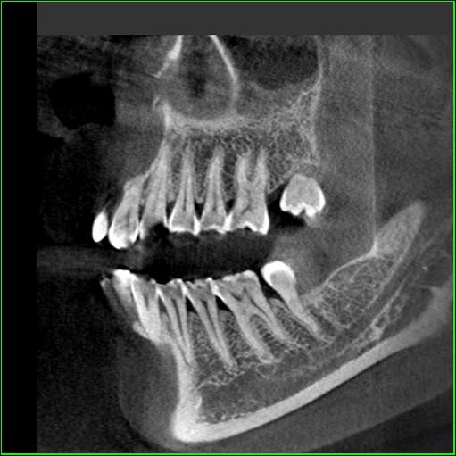
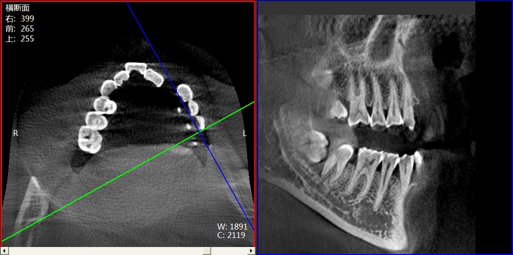
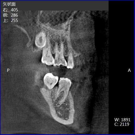
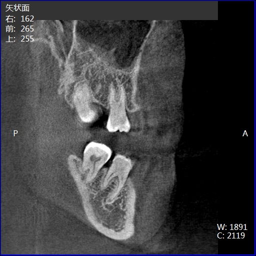
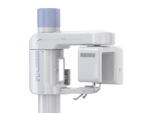
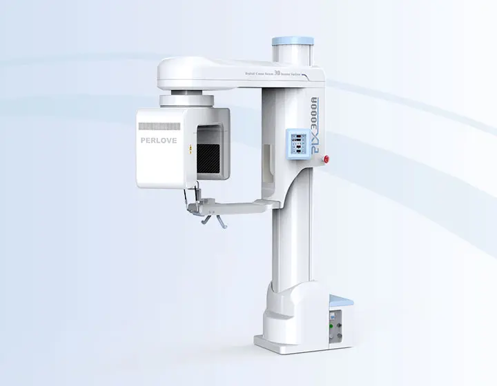
Great Quality Images PLX3000A
| X-Ray SourceAdopt pulse exposure mode, real exposure time is 4s during 14s scanning, reduce the x-ray dose a lot, green and safety. |
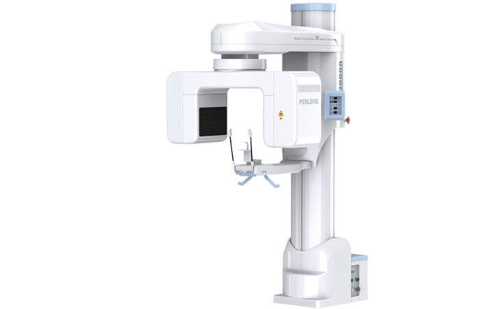
Imaging System
|
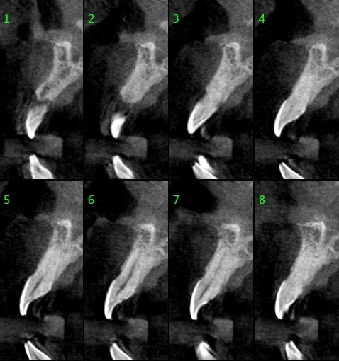
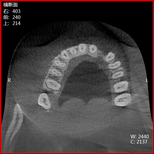
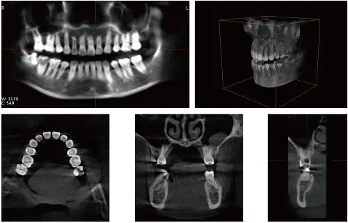
Great Quality ImagesAdopt 3D reconstruction algorithm. |
Operating Interface
|
Digital network seamless linkDicom 3.0 network interface for integration with clinical network, PACS and RIS. |

Adopt pulse exposure mode, real exposure time is 4s during 14s scanning, reduce the x-ray dose a lot, green and safety.
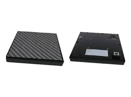
Adopt Thales Flat Panel Detector
High Resolution, Small Distortion, Brightness Uniformity
Effectve size: 12cmX15cm flat panel detector


Touch Screen
Freindly interface, easy and convenient
Ergomomics Design
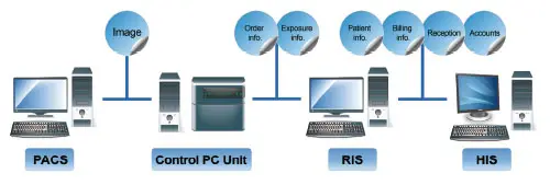
Dicom 3.0 network interface for integration with clinical network, PACS