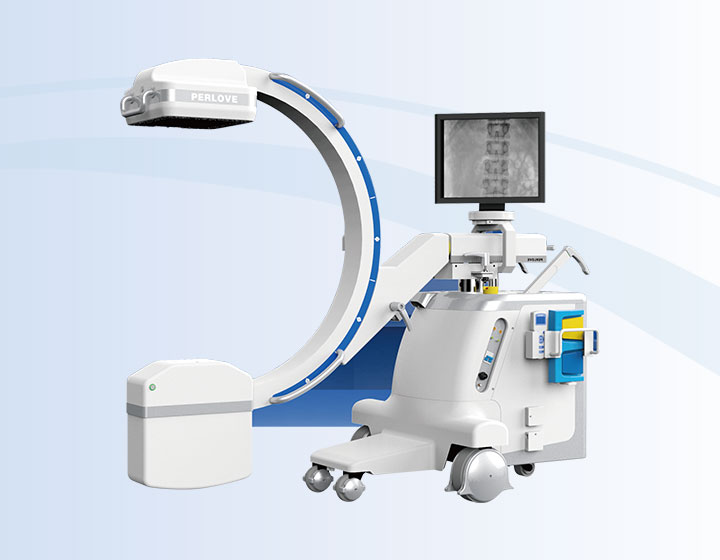
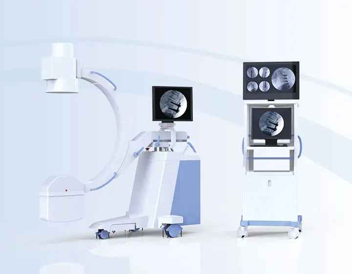
Mobile Digital FPD C-arm System
Dynamic FPD Technology Brilliant image quality
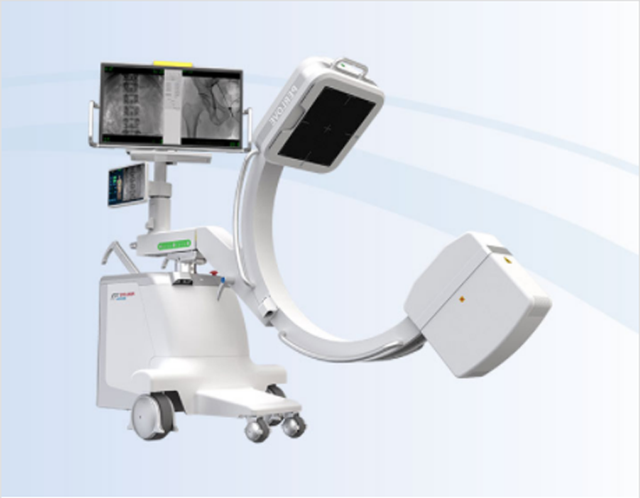
Compared to the conventional 9” image intensifier, a 9” x 9” flat panel detector is able to expand the FoV by 22%, providing more perspective for clinical diagnosis.
Higher signal coversion efficiency of FPD ensures higher image quality and lower dose.
|
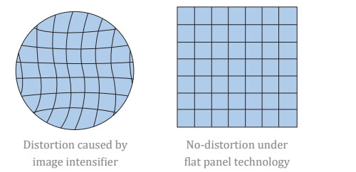
Eliminating distorted image caused by electron beam deflection, presenting actual vision of anatomical structure.
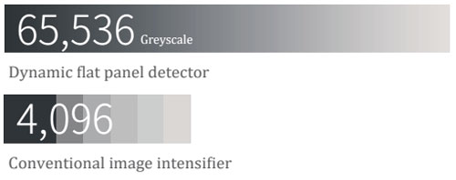
16 bits depth determines maximum 65536 greyscale value, providing HD resolution for revealing more anatomical details.
|
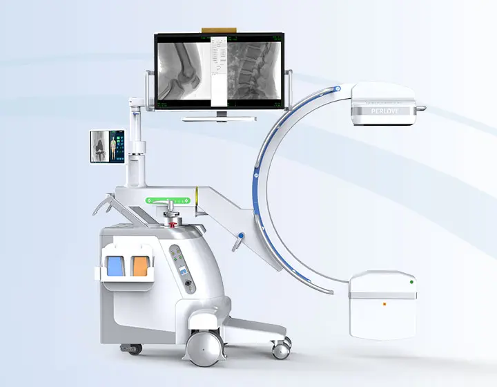
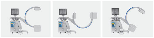
Three simple steps to achieve clear imageOn/off push button switch intergrated One button press to switch on/off the console, simplifies overall workflow, saving preparation time. Two-way laser targeting Built-in laser positioning system at both X-ray tube and flat panel detector. Precisely lesion targeting in different C-arm position, improving diagnostic efficiency. Flexible C-arm movement Exquisite C-arm design ensures maximum flexibility: 200mm horizontal movement, ±15° swivel range, 135° orbital movement and 400mm motorized elevation.
| Intelligent image processing Automatically adjust and optimize exposure parameters, simplify operation and enjoy easy examination. Touchscreen display User-friendly touchscreen interface makes every step
|
Apparent simplicity with outstanding user experienceHigh DQE and greyscale value, low noise level, giving excellent image with crystal clarity.
Specialized for pediatrics and dose sensitive patients.
| Large heat capacity and fast heat dissipation ensures working continuously.
Remote control C-arm movement and exposure parameter adjustment.
|
Non-cable connection design prevent potential stumble accident.
No exposure when button locked, avoiding radiation caused by misoperation.
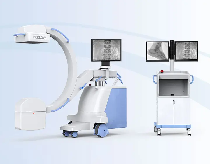
| Large angle rotatable monitors provide multiple observation perspectives.
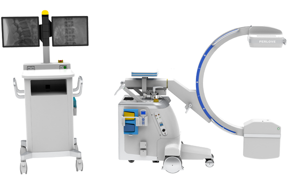
Lightweight slim design, flexible
|
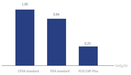
Low dose management Multiple protectionLeakage radiation in the loading sate of X-ray tube is far more lower than CFDA and FDA standard, providing better protection from the X-ray source.
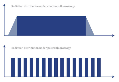
Real time DAP meter monitoring is available. Measuring actual dose level and displaying on the console. 110kHz monoblock high frequency high power generator ensurse high quality X-ray, reducing soft X-ray level and radiation dose. | X-ray generated in high frequency pulse wave can reduce 50% of radiation dose compared with a continous flouroscopy. This particular technology has a positive effect on weakening the motion artifact.
Allowing radiologist to preview and to adjust exposure area before exposure, reducing unnecessary radiation absorption. Decreasing radiation level by absorbing soft X-ray. Ensuring high quality image for precise |
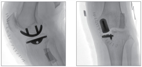
Multidisciplinary use Versatile surgical solutionDescription: Patient has suffered from intractable pain caused by oseoarthritis and underwent incurable conservative treatment. During the operation, the image clearly showed the shape of bone structure and joint surface, which helped the doctor to implant the prosthesis accurately
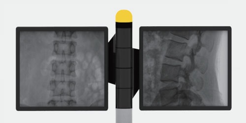
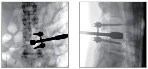
Description: Lumbar disc herniation, frequent pain in lower limbs. In order to decompress the vertebrae, pedicle screws are accurately implanted into the lumbar spines by the image guidance of lumbar AP and lateral view. |
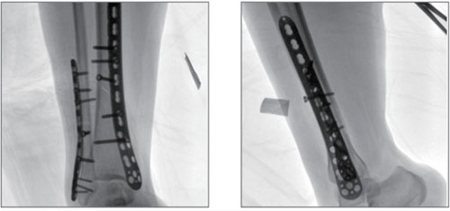
Description: Distal tibia and fibula oblique fracture caused by car accident. By the AP/ lateral view image of tibia and fibula, compression plates are accurately implanted into the bones to achieve stability.
|

Compared to the conventional 9” image intensifier, a 9” x 9” flat panel detector is able to expand the FoV by 22%, providing more perspective for clinical diagnosis.


Higher signal coversion efficiency of FPD ensures higher image quality and lower dose.
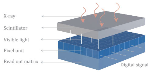
Eliminating distorted image caused by electron beam deflection, presenting actual vision of anatomical structure.

16 bits depth determines maximum 65536 greyscale value, providing HD resolution for revealing more anatomical details.

On/off push button switch intergrated
One button press to switch on/off the console, simplifies overall workflow, saving preparation time.
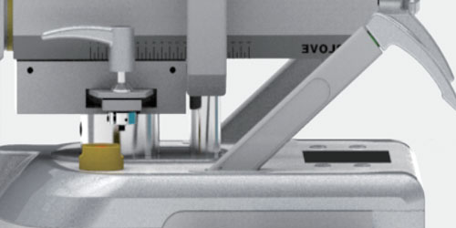
Two-way laser targeting
Built-in laser positioning system at both X-ray tube and flat panel detector. Precisely lesion targeting in different C-arm position, improving diagnostic efficiency.
Flexible C-arm movement
Exquisite C-arm design ensures maximum flexibility: 200mm horizontal movement, ±15° swivel range, 135° orbital movement and 400mm motorized elevation.
Offering easy patient coverage at different position
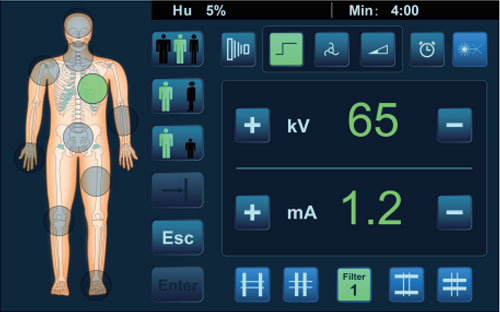
Intelligent image processing
Automatically adjust and optimize exposure parameters, simplify operation and enjoy easy examination.
Touchscreen display
User-friendly touchscreen interface makes every step
of operation easy and clear.

High DQE and greyscale value, low noise level, giving excellent image with crystal clarity.
Specialized for pediatrics and dose sensitive patients.
Remove the grid to reduce radiation absorption.
Large heat capacity and fast heat dissipation ensures working continuously.

Remote control C-arm movement and exposure parameter adjustment.
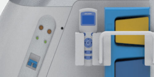
Non-cable connection design prevent potential stumble accident.

No exposure when button locked, avoiding radiation caused by misoperation.
Large angle rotatable monitors provide multiple observation perspectives.

Lightweight slim design, flexible
maneuverablility.

Leakage radiation in the loading sate of X-ray tube is far more lower than CFDA and FDA standard, providing better protection from the X-ray source.
Real time DAP meter monitoring is available. Measuring actual dose level and displaying on the console.
110kHz monoblock high frequency high power generator ensurse high quality X-ray, reducing soft X-ray level and radiation dose.
X-ray generated in high frequency pulse wave can reduce 50% of radiation dose compared with a continous flouroscopy. This particular technology has a positive effect on weakening the motion artifact.
Allowing radiologist to preview and to adjust exposure area before exposure, reducing unnecessary radiation absorption.
Decreasing radiation level by absorbing soft X-ray. Ensuring high quality image for precise
diagnosis and treatment.
Description: Patient has suffered from intractable pain caused by oseoarthritis and underwent incurable conservative treatment.
During the operation, the image clearly showed the shape of bone structure and joint surface, which helped the doctor to implant the prosthesis accurately
Description: Lumbar disc herniation, frequent pain in lower limbs.
In order to decompress the vertebrae, pedicle screws are accurately implanted into the lumbar spines by the image guidance of lumbar AP and lateral view.

Description: Distal tibia and fibula oblique fracture caused by car accident.
By the AP/ lateral view image of tibia and fibula, compression plates are accurately implanted into the bones to achieve stability.