01 A right operation table
02 Patient positioning
03 Use exposure protection
04 Set up the C-arm
05 Positioning in the isocenter with laser
06 Parameter setting
07 Collision check
08 Start the scan with footswitch
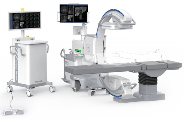
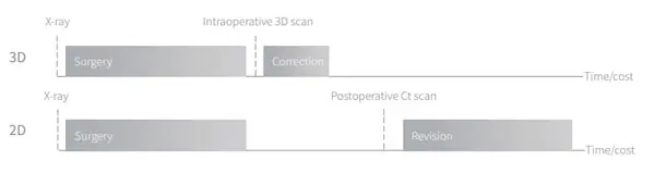
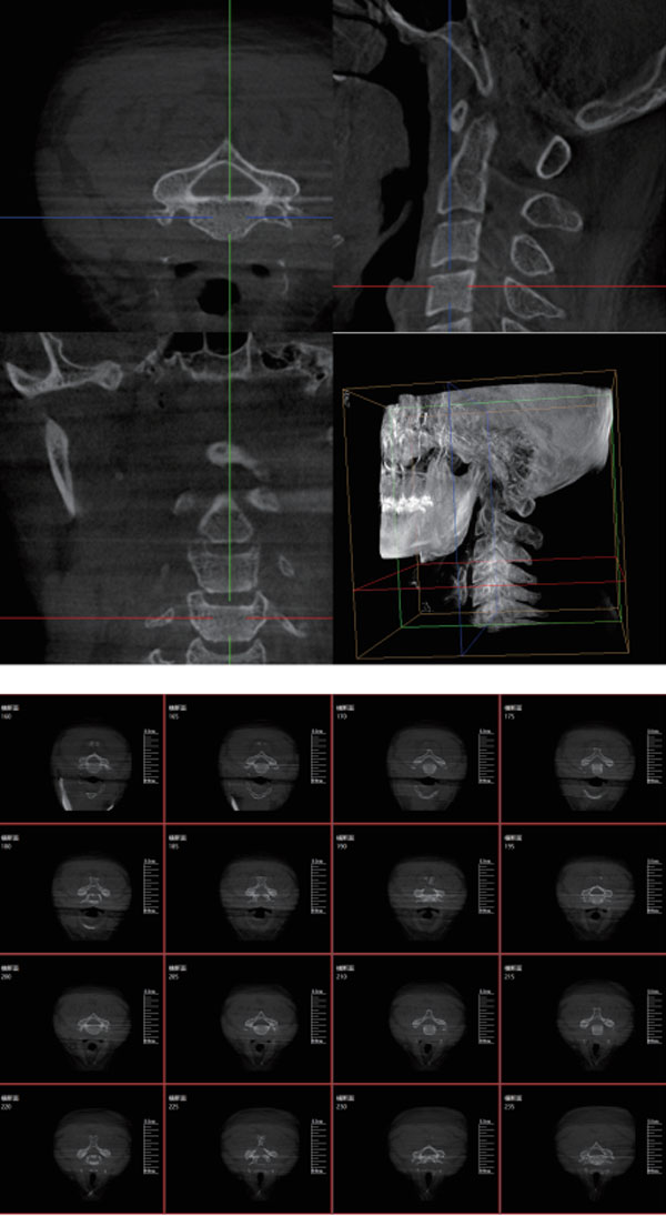
Intraoperative 3D imaging and CT-like imaging provides precise information from every angle during the surgical procedures—-pinpoint anatomical structures, implants and screws more confidently.
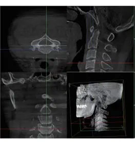
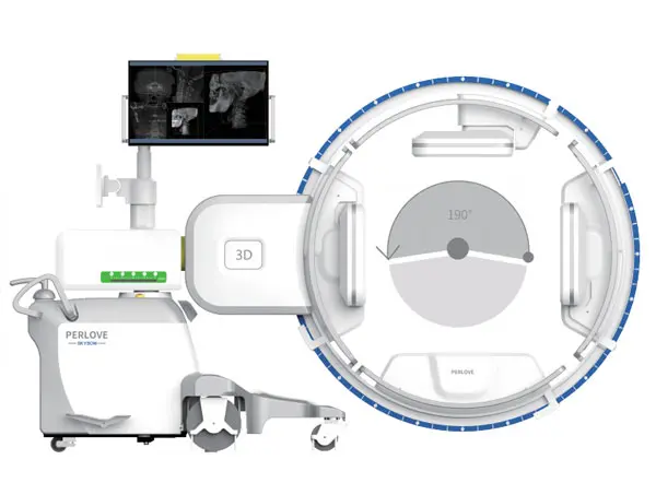
Delivers a 3D image covering a volume of a vertical cylinder more information would be seen in one volume:
● Seven cervical vertebrae
● Seven thoracic vertebrae
● Five lumbar vertebra
● Bilateral iliosacral joints
● Femur head and unilateral pelvis
Intuitive intraoperative 3D evaluation avoids unnecessary postoperative CT scans and corrective surgery, saving time and costs.
With iso-centric scan technology, orbital movement in a motorized 3D scan from any direction giving you complete, highly accurate 3D information in outstanding quality.
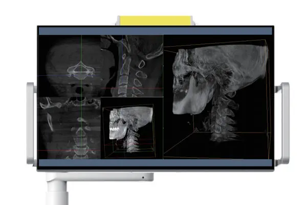
wide medical monitor showing images with high brightness and high contrast.
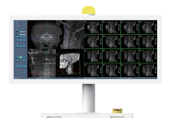
integrated monitor presents real-time images for easy image reading, relieving the burden of memory.
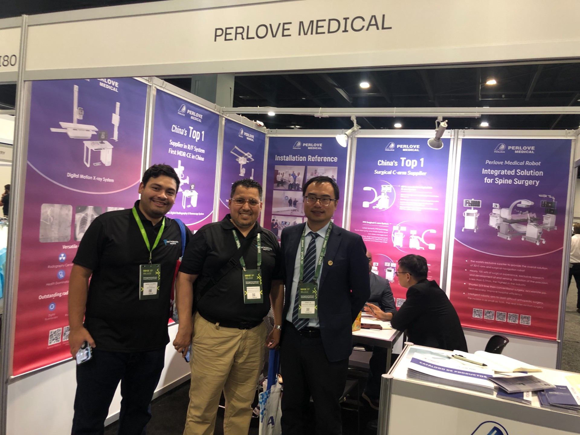
🌍 WHX Miami 2026 – Exhibition Wrap-Up
Read More »Perlove Medical to Exhibit at WHX Miami — See You in Miami!
Read More »A Decade of Dedication, Echoing in the Market Again | Perlove Medical’s C-arms Deployed in Three Major Regional Hospitals in Bangladesh
Read More »2026 Innovate Together, Grow Further丨Perlove Medical International Distributor Promotion Conference
Read More »PERLOVE MEDICAL at ECR 2026 | Advancing Radiology Innovation in Vienna
Read More »Perlove Medical at ECR 2026: Showcasing Cutting-Edge Imaging Solutions in Vienna
Read More »