2025-08-21 16:48:25 Views:1480
Due to academic milestones such as high school entrance examination, adolescent patients with scoliosis often find it difficult to allocate several weeks for postoperative recovery during the school year. As a result, summer vacation has become the “golden window” corrective spinal surgery in this population. From July to August, Children's Hospital of Nanjing Medical University used orthopedic surgical robots to perform scoliosis corrective surgery on several female patients aged 12 to 15, helping the girls regain their upright posture.
01 Health Education | Caring for Adolescent Spinal Health
Scoliosis is a complex 3D spinal deformitye. According to the Scoliosis Research Society (SRS), it is defined as a Cobb angle of ≥10° measured on standing anteroposterior full-spine radiographs. Idiopathic scoliosis, which accounts for approximately 80% of all scoliosis cases, has no clearly identified etiology. Among these, Adolescent Idiopathic Scoliosis (AIS) is the most common type, occurring in children aged 10 to 18 years, with a prevalence ranging from 1.5% to 3.0%.
AIS affects adolescents’appearance, causes back pain and psychological disorders, and in severe cases may lead to respiratory dysfunction and impaired mobility. This creates a heavy burden on both families and society. Therefore, early prevention, diagnosis, and precise treatment of adolescent scoliosis are particularly important.
For adolescents with severe scoliosis who meet surgical indications, precise surgical intervention can not only effectively correct spinal deformities and restore the normal physiological curvature and biomechanical balance of the spine, but also provide strong support for psychological health. This helps them integrate better into society and grow up healthily and confidently.
02 Case Sharing | Robot-Assisted Spinal Deformity Surgery
Spinal deformity surgery involves long-segment fixation and the precise placement of multiples pedicle screws. This is especially challenging in areas with severe rotational misalignment and vertebral deformities, where extremely high accuracy in 3D spatial positioning is required. It is one of the most technically demanding procedures in spinal surgery.
At Children's Hospital of Nanjing Medical Universityl, robot-assisted screw placement has enabled precise control of bleeding and operative time in anatomical areas, balancing both efficiency and safety. This approach has provided a new paradigm for open surgery in adolescent idiopathic scoliosis (AIS).
Case 1
Patient: 12-year-old female
Surgical site: T4-L3
More accurate, more stable, lasting care
The long-term stability of internal fixation directly determines the maintenance of correction and the quality of fusion during the adolescent skeletal development period. With sub-millimeter precision, robotic navigation enables one-time screw placement, ensuring that the screw trajectory is positioned in the thickest part of the cortical bone to maximize screw load-bearing capacity. At the same time, it minimizes cancellous bone damage caused by repeated adjustments, thereby reducing the risk of screw loosening and pseudoarthrosis.

3D C-arm intraoperative fluoroscopy + orthopedic surgical robot path navigation

The doctor completed the screw placement under the guidance of robotic planning.
Precise screw trajectory planning and stable fixation create an optimal biomechanical environment for intervertebral fusion. Combined with adolescents' vigorous bone healing potential, this effectively promotes early bony fusion and significantly improves long-term prognosis.

Case 2
Patient: 14-year-old female
Surgical site: T2–L4
Reduced blood loss, making it safer.
In scoliosis correction surgery, to ensure the accuracy of screw placement, repeated adjustments and multiple fluoroscopic confirmations are usually required, which carries the risks of prolonged operation time and increased blood loss. In robot-assisted surgery, doctors can complete the planning of screw placement for multiple vertebrae at one time based on intraoperative 3D images and quickly achieve precise screw placement. This eliminates the time spent on repeated adjustments, avoids prolonged manipulation in high-risk anatomical areas, and can effectively control blood loss in high-risk regions
.
3D C-arm for intraoperative 3D image acquisition.
The optimal path planning for the insertion angle and depth of multiple screws is completed at the robotic workstation.
Precise positioning of screw insertion under the guidance of the robotic arm
Image verification shows good fusion effect.
Case 3
Patient: 15-year-old female
Surgical site: T5-L3
3D imaging for precise assurance
The upper thoracic vertebrae from T5 to T7 exhibit severe spinal rotation and vertebral distortion, requiring simultaneous reduction in the coronal, sagittal, and axial planes, demands extremely high precision in 3D spatial positioning. Based on the clear 3D images provided by the 3D C-arm, the dsurgeon precisely planned the trajectory, angle, and depth of screw placement in the robotic navigation system. With sub-millimeter navigation accuracy and excellent stability, the robotic arm guided the surgeon to accurately insert the screws into the predetermined target points in one attempt.


Accurate screw placement for optimal correction
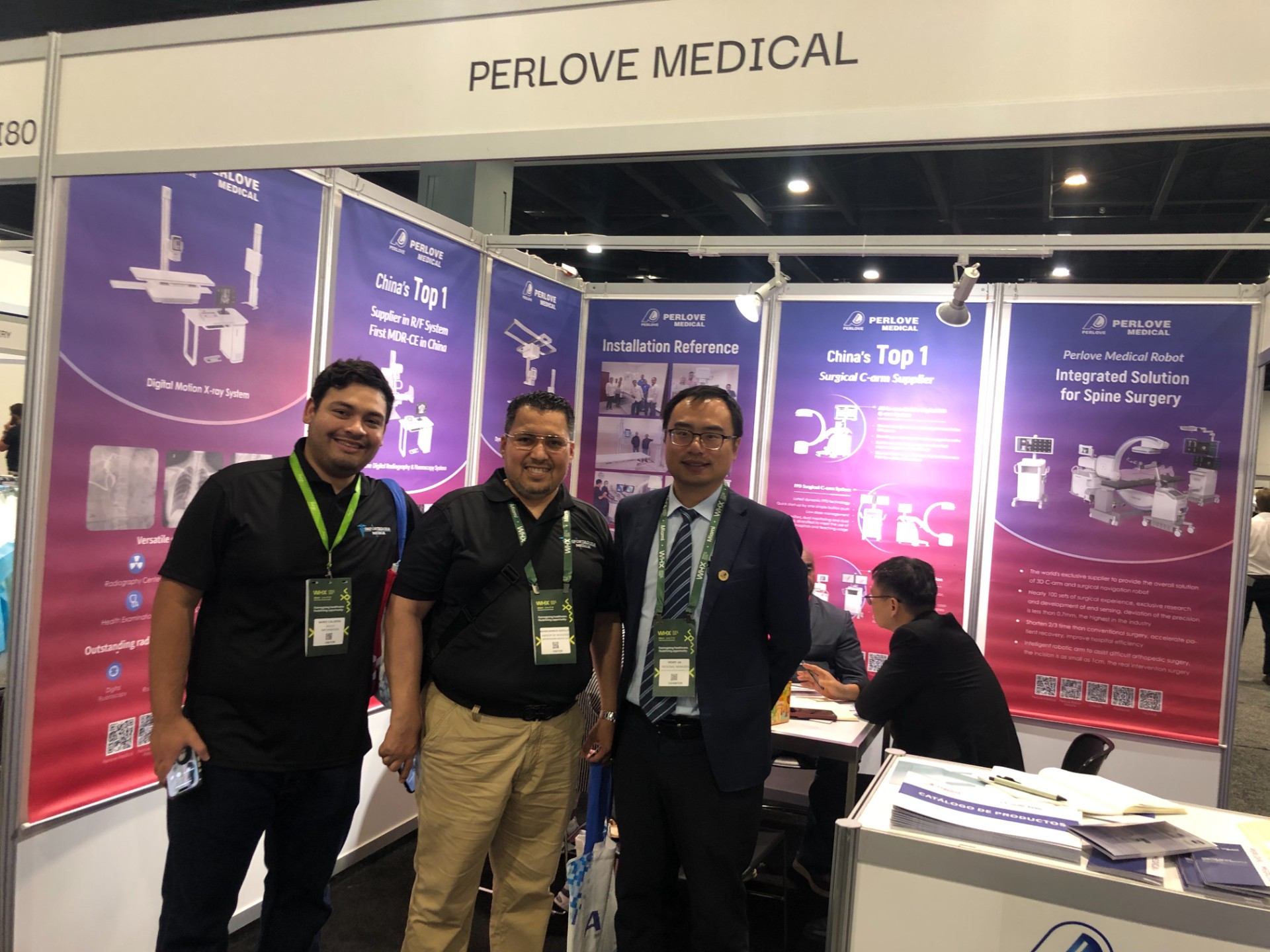
🌍 WHX Miami 2026 – Exhibition Wrap-Up
Read More »Perlove Medical to Exhibit at WHX Miami — See You in Miami!
Read More »A Decade of Dedication, Echoing in the Market Again | Perlove Medical’s C-arms Deployed in Three Major Regional Hospitals in Bangladesh
Read More »2026 Innovate Together, Grow Further丨Perlove Medical International Distributor Promotion Conference
Read More »PERLOVE MEDICAL at ECR 2026 | Advancing Radiology Innovation in Vienna
Read More »Perlove Medical at ECR 2026: Showcasing Cutting-Edge Imaging Solutions in Vienna
Read More »