Since the introduction of the Perlove Medical orthopedic surgical robot,the Affiliated Traditional Chinese Medicine Hospital of Chongqing Three Gorges Medical College has performed over 200 robotic-assisted spinal surgeries, including MIS-TLIF for lumbar disc herniation, lumbar spinal stenosis, and lumbar spondylolisthesis, percutaneous minimally invasive pedicle screw fixation for thoracolumbar fractures, PKP/PVP for osteoporotic vertebral fractures, and scoliosis correction. These advanced procedures have significantly enhanced the precision, efficiency, and safety of minimally invasive spinal treatment.
In the field of spinal surgery, the development of minimally invasive technology has always focused on the core goals of "smaller incisions, better outcomes, and faster recovery." In recent years, robot-assisted minimally invasive transforaminal lumbar interbody fusion (MIS-TLIF) has gradually become an important surgical approach for treating degenerative lumbar diseases (such as lumbar disc herniation, lumbar spinal stenosis, and lumbar spondylolisthesis) due to its high precision and strong safety.

The technology deeply integrates the robot's "precise operation" with the "minimally invasive concept" of MIS-TLIF, combined with intraoperative 3D imaging and real-time navigation systems, significantly improving the accuracy and safety of the surgery, providing patients with a completely new treatment experience.
MIS-TLIF (Minimally Invasive Transforaminal Lumbar Interbody Fusion) is a minimally invasive technique developed based on traditional open TLIF surgery. Compared with traditional open surgery, this approach accesses the surgical area through a small incision or tubular retractors, minimizing extensive dissection and retraction of the back muscles. As a result, it significantly reduces intraoperative blood loss, postoperative pain, muscle atrophy, and other complications, while shortening the patient's hospital stay and recovery time.
However, the MIS-TLIF procedure has limited operating space and involves complex anatomical structures (including critical tissues such as nerves, blood vessels, and the dural sac), which requires high precision from the surgeon. The introduction of robot-assisted technology addresses this core challenge.
Patient: 77 years old, female
Preoperative Diagnosis: Lumbar spinal stenosis, lumbar instability
Surgical Plan: Robot-assisted MIS-TLIF surgery, with decompression first followed by screw placement.

During the procedure, Perlove Medical's 3D C-armperformed a 3D scan of the lumbar spine., and the 3D images were then imported into the robotic work system. The surgeon used the system's software to observe the patient's lumbar anatomy (intervertebral disc height, vertebral body size) and identified the diseased segment (L4-L5). A comprehensive surgical plan was then developed, defining the optimal trajectory for screw placement (including angle, depth, and diameter) and the optimal size and position for the interbody cage, ensuring the plan was customized to the patient's individual anatomy.


Following the preplanned trajectory for screw placement, the surgeon made two small incisions (approximately 2–3 cm) on both sides of the affected spinal segment on the patient’s back. Through these incisions, dilators were inserted to gradually separate the subcutaneous tissue and muscles without cutting them. This process ultimately created a working channel with a diameter of only 3–4 cm, allowing precise access to the surgical segment. This technique maximizes the preservation of lumbar muscles and minimizes the risk of postoperative pain and functional weakening.

The robotic arm moved to the planned entry points, with its guide sleeve perfectly aligned with the pre-planned trajectory. The surgeon then drilled four Kirschner wires through this guide.
After intraoperative verification confirmed accurate positioning, four pedicle screws were placed. Compared to the traditional freehand technique, robot assistance eliminates deviations caused by reliance on subjective tactile feedback, significantly enhancing the accuracy of screw placement.


After screw placement was completed, the surgeon proceeded through the working channel to access the foraminal region. Using specialized minimally invasive instruments, the pathological disc tissue was removed to decompress the nerve root. Next, the preselected interbody fusion cage was implanted into the intervertebral space via the channel to restore disc height and reconstruct lumbar stability, creating an optimal environment for interbody fusion.
Finally, the surgeon contoured the connecting rods and introduced them through the working channel. The rods were then secured to the previously placed pedicle screws, further reinforcing lumbar stability. After confirming the correct position of the construct, the incision was closed in layers, completing the procedure.

New Breakthroughs in Surgical Efficiency and Fusion Outcomes

专家介绍Yang Dong — Director of Orthopedics Department I
Affiliated Traditional Chinese Medicine Hospital of Chongqing Three Gorges Medical College
Specializing in minimally invasive surgical treatments, Dr. Yang is highly experienced in endoscopic procedures for cervical spondylosis, lumbar disc herniation, and lumbar spinal stenosis through the intervertebral foramen. He is also skilled in minimally invasive channel surgeries for lumbar instability, as well as treatments for spinal fractures and severe osteoporosis. Dr. Yang was among the first in Northeast Chongqing to perform robot-assisted minimally invasive spine surgeries, enabling highly precise minimally invasive interventions for spinal disorders.
Robot-assisted screw placement and fusion cage implantation ensure a stable internal fixation system and even distribution of forces within the intervertebral space. This creates an optimal biomechanical environment for interbody fusion, significantly improving the postoperative fusion rate while reducing complications such as pseudoarthrosis formation and screw loosening.
With the combination of a 3D C-arm and robotic navigation system, a single scan can capture three-dimensional images of the surgical area, enabling the planning of multiple screw placements at once. This minimizes the time surgeons spend repeatedly confirming screw positions, eliminating the need for multiple fluoroscopic checks. Compared to traditional MIS-TLIF procedures, this approach shortens the overall operation time and improves surgical efficiency.
Robot-assisted MIS-TLIF surgery represents a breakthrough for the Department of Spine Surgery at the Affiliated Traditional Chinese Medicine Hospital of Chongqing Three Gorges Medical College, overcoming the “precision limitations” of conventional minimally invasive techniques. It drives lumbar fusion surgery toward smaller incisions, better outcomes, and faster recovery.
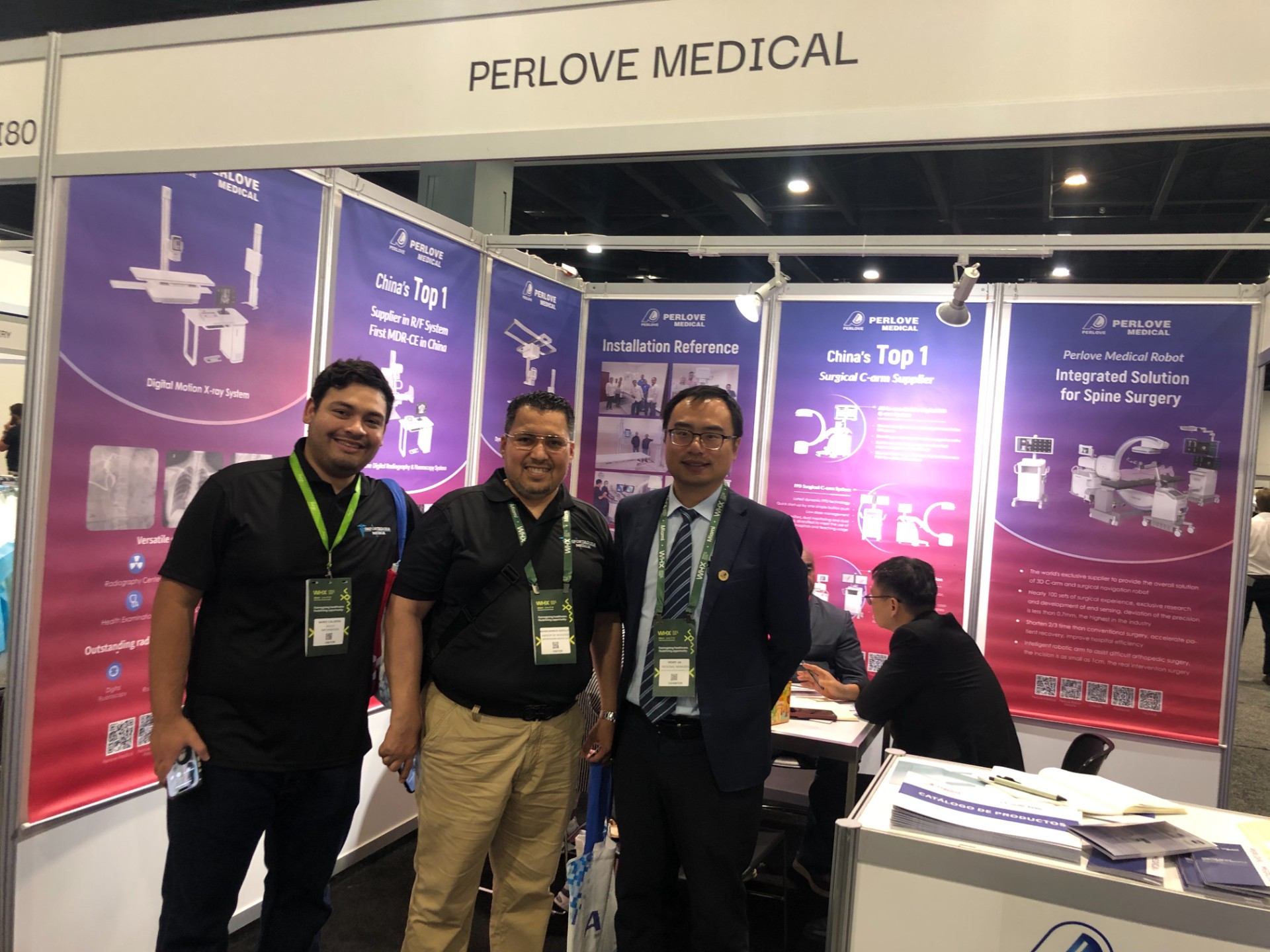
🌍 WHX Miami 2026 – Exhibition Wrap-Up
Read More »Perlove Medical to Exhibit at WHX Miami — See You in Miami!
Read More »A Decade of Dedication, Echoing in the Market Again | Perlove Medical’s C-arms Deployed in Three Major Regional Hospitals in Bangladesh
Read More »2026 Innovate Together, Grow Further丨Perlove Medical International Distributor Promotion Conference
Read More »PERLOVE MEDICAL at ECR 2026 | Advancing Radiology Innovation in Vienna
Read More »Perlove Medical at ECR 2026: Showcasing Cutting-Edge Imaging Solutions in Vienna
Read More »