2025-12-15 17:03:36 Views:1493
The trigeminal nerve is the primary somatosensory nerve of the face and consists of three branches: the ophthalmic branch, the maxillary branch, and the mandibular branch, which are responsible for sensory transmission in the forehead, midface (including the cheek), and lower jaw regions, respectively. When the trigeminal nerve is subjected to vascular compression, neoplastic invasion, or inflammatory stimulation, it can easily lead to paroxysms of excruciating pain. This pain is typically described as electric shock-like (lancinating), stabbing, or burning in nature, and is often triggered by everyday activities such as washing the face, brushing teeth, or chewing, thereby severely affecting the patient’s quality of life.
In the minimally invasive interventional treatment of trigeminal neuralgia, radiofrequency thermocoagulation has become one of the most widely used clinical techniques due to its simple procedure, reliable therapeutic efficacy, and the feasibility of repeat treatment in cases of recurrence. Common puncture approaches include the Hartel anterior approach, the submandibular–foramen ovale approach, and the lateral approach. Through these routes, high-frequency electrical current is delivered via an electrode to selectively ablate the pain-transmitting sensory fibers within the trigeminal ganglion, while preserving motor fibers and tactile sensory fibers.
The foramen ovale is an oval-shaped opening located in the greater wing of the sphenoid bone at the skull base, with a diameter of only 3–5 mm. It serves as a critical passage for the mandibular branch of the trigeminal nerve, the lesser petrosal nerve, and related vasculature, and is also the only “gateway” through which a radiofrequency electrode can percutaneously access the intracranial trigeminal ganglion.
Due to its deep anatomical location and significant inter-individual variability, even a minor deviation in trajectory can result in failure to cannulate the target neural structure or, more critically, misplacement into adjacent vital tissues. Therefore, precise localization of the foramen ovale represents the most critical challenge of the procedure. Accurate targeting can reduce the number of puncture attempts, shorten operative time, and lower both intraoperative patient discomfort and radiation exposure. Moreover, precise localization helps preserve normal nerve function to the greatest extent, reducing the incidence of postoperative complications such as facial numbness (hypesthesia) or dysesthesia, thereby improving patients’ quality of life and lowering the recurrence rate.

Schematic Diagram of the Foramen Ovale
With advantages such as flexible mobility and 3D precise imaging, the mobile flat-panel 3D C-arm from Perlove Medical achieves full-process imaging support from puncture positioning to ablation effect verification through 3D scanning, bringing safer and more efficient treatment experiences to patients.

3D Image-Guided Localization of the Foramen Ovale
The patient is placed in a supine position. The C-arm is flexibly adjusted to the optimal scanning angle to perform rapid 3D scanning of the foramen ovale region at the skull base. The device automatically reconstructs a 3D anatomical model of the foramen ovale and the surrounding skull, clearly displaying the spatial location, aperture size of the foramen ovale as well as its adjacency to surrounding structures, thereby providing an accurate anatomical basis for puncture path planning.

Based on the 3D images, the doctor plans the optimal puncture path to ensure precise alignment between the puncture direction and the foramen ovale, minimizing puncture deviations.

Electrical Stimulation Test
After the puncture needle reaches the target area, it is connected to a radiofrequency generator for electrical stimulation testing, while the patient’s facial responses are observed to confirm the precise positioning of the electrode tip at the root of the specific trigeminal branch responsible for the pain. Once positioning is completed, anesthetic and sedative drugs are administered intravenously to induce a controlled sleep state in the patient, followed by radiofrequency ablation. After the radiofrequency ablation treatment, the patient is allowed to awaken, the puncture wound is dressed with an adhesive bandage, and the surgery is completed.
Mobile Design for Diverse Scenarios: Compared with fixed-installation DSA equipment, the mobile flat-panel 3D C-Arm from Perlove Medical features a compact size and flexible mobility. It can be adjusted in position according to surgical needs, adapting to operating room layouts of different specifications. Particularly suitable for emergency settings or multi-department collaborative scenarios, it enhances equipment utilization efficiency.
With rapid 3D scanning and reconstruction technology, the device can clearly visualize the 3D spatial structure of the foramen ovale and trigeminal nerve. It addresses the problem that traditional 2D fluoroscopy struggles to accurately assess depth and critical spatial relationships with adjacent anatomy, thus providing reliable support for puncture path planning in complex clinical cases.
The 3D scanning and reconstruction process is fast and efficient, which can significantly shorten preoperative preparation time. Meanwhile, precise localization reduces the number of intraoperative adjustments, streamlines overall treatment workflow, and alleviates the surgical burden on patients.

A Decade of Dedication, Echoing in the Market Again | Perlove Medical’s C-arms Deployed in Three Major Regional Hospitals in Bangladesh
Read More »2026 Innovate Together, Grow Further丨Perlove Medical International Distributor Promotion Conference
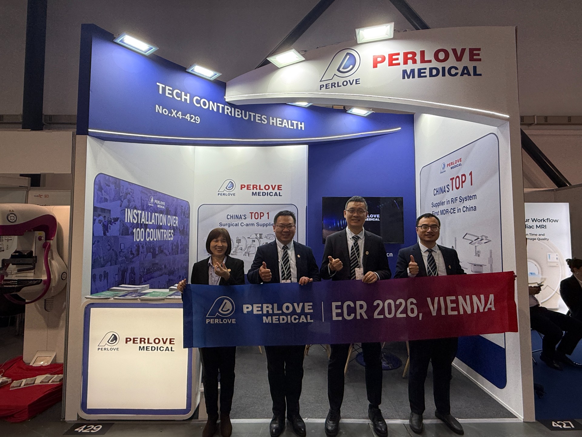
Read More »PERLOVE MEDICAL at ECR 2026 | Advancing Radiology Innovation in Vienna
Read More »Perlove Medical at ECR 2026: Showcasing Cutting-Edge Imaging Solutions in Vienna
Read More »🌍 Elevating Global Healthcare | WHX Dubai 2026 Successfully Concluded
Read More »Join Us at ARAB HEALTH 2026 | See You in Dubai
Read More »