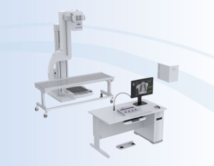
Full-Field Dynamic Imaging Full-Field Dynamic Imaging

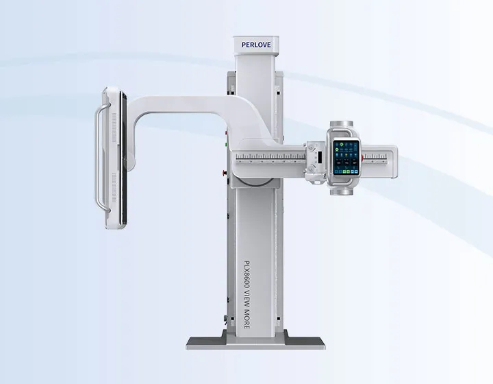
Single-Panel DRPLX8600 Series Applicable Departments: Orthopedics,Trauma, Spinal Orthopedics, Interventional Radiology, etc.  | Dynamic/Static ImagingSingle PanelPrecise data Full-field Maximum 100kw Fast exposure Dynamic ImagingLarge-field dynamic FPD |
Breakthrough in Field of ViewFull-Field supine lmaging | Large-Field Dynamic Flat PanelSeamless lmaging Without Stitching |
Premium Imaging SolutionMaximum 100kw - superior X-ray qualityAdapted for diverse body typesThe PLX8600 series combines precision engineering with advanced imaging technology, incorporating a proprietary high-power generator for superior X-ray quality. The system features an imported high-capacity X-ray tube and proprietary image processing algorithms to deliver high-quality diagnostic images with minimal latency, reducing missed diagnoses or diagnostic errors.
| Effortless Positioning, Instant lmaging
Highly integrated gantry |
Meticulous CraftsmanshipThe system features a highly integrated architecture with a proprietary high-voltage generator and imported x-ray tube. It is equipped with the fuli-field flat panel detector to deliver both static and dynamic imaging in upright and supine positions. The system adopts proprietary image processing algorithms during image acquisition, ensuring diagnostic accuracy and reliability for every examination. Quality for an Enduring BrandCraftsmanship is revealed in the details. Quality forges an evergreen brand. Precision Engineering and Advanced Imaging | Precise dose measurement Fast exposure
Compact, space-saving
X-ray quality assurance
Superior image quality
|
Broad Clinical ApplicationsAdolescent Scoliosis ScreeningSingle-Shot Imaging for Full Spine Precise Cobb Angle Measurement | Lower Radiation Dose
High-Quality Clinical lmages for Adolescent Scoliosis Adolescent Spinal Health Precision Spinal Care for Healthier Futures |
Broad Clinical ApplicationsFull-Lower-Limb DiagnosticsSingle-Shot lmaging for Full Lower Limbs Accurate Mechanical Axis Measurement |
Clinicallmages for Pre-/Post-Op Full-Lower-Limb Trauma Precision & Efficiency for Clinical Applications Each surgical procedure embodies a vital collaboration between surgeons, patients and their families, delivering precision treatment to restore mobility and quality of life through Perlove’s clinically-proven, efficiency-optimized therapeutic approaches. |
PLX8600 Series
Applicable Departments: Orthopedics,Trauma, Spinal Orthopedics, Interventional Radiology, etc.
Clinical Applications: Full-spine imaging, full-lower-limb radiography, complex trauma cases, joint replacement and reconstruction etc.
Precise data
Efficient positioning
Low radiation dose
Full-field
upright/supine imaging
Maximum 100kw
superior X-ray quality
Adapted for diverse body types
Fast exposure
lnstant imaging
Large-field dynamic FPD
Max.FOV 0.43m×1.2m
All-in-one system
Cost-efficient
Single Shot, Full Coverage
From feet to anterior superior iliac spine(ASIlS), all ages
Surgical Accuracy
Measures limb alignment length and angular parameters for lower limb deformity correction, high tibial osteotomy (HTO), and total knee arthroplasty (TKA)
Full-field supine lmaging, Critical Care
Optimized for mobility-limited patients, less repositioning in emergencies
Single-shot Acquisition
No stitching artifacts, crystal-clear results
Rapid Response
Minimal exposure time, higher diagnostic speed

Clinicallmages for Pre-/Post-Op Full-Lower-Limb Trauma
Ultra-Wide FOV
0.43m×1.2m (3× conventional detectors)
Full Spine/Lower Limb
Single-shot capture, no stitching
Less Dose
Compared with stitched DR systems
Better Alignment
Eliminates density variations and junction misalignment
Enhanced Workflow
Faster, more accurate diagnoses
All-in-One lmaging
Radiography, fluoroscopy and spot imaging
Large-Field Dynamics
High-quality multi-angle visualization
Smart Capture
Critical frame acquisition reduces repeat exposures
Specialized Application Excellence
ldeal for articular kinematics, Gl contrast studies, full-spine assessments, complex positioning guidance, etc.

The PLX8600 series combines precision engineering with advanced imaging technology, incorporating a proprietary high-power generator for superior X-ray quality. The system features an imported high-capacity X-ray tube and proprietary image processing algorithms to deliver high-quality diagnostic images with minimal latency, reducing missed diagnoses or diagnostic errors.


The PLX8600 series incorporates a fully integrated generator-gantry system with a unified design. This advanced configuration operates without ceiling tracks while requiring minimal vertical clearance for installation.The space-efficient design enables quick deployment in standard radiology departments without structural modifications.
The system features a highly integrated architecture with a proprietary high-voltage generator and imported x-ray tube. It is equipped with the fuli-field flat panel detector to deliver both static and dynamic imaging in upright and supine positions. The system adopts proprietary image processing algorithms during image acquisition, ensuring diagnostic accuracy and reliability for every examination.
Craftsmanship is revealed in the details. Quality forges an evergreen brand.
Full-field imaging, efficient diagnosis

SID-specific design

Precise dose measurement
Fast exposure
without adjusting parameters

Compact, space-saving

X-ray quality assurance

Superior image quality

Single-Shot Imaging for Full Spine
The system’s ultra-large full field of view enables single-shot, non-stitched imaging with enhanced precision, allowing full-spine visualization in a single exposure for spinal orthopedic procedures and other surgical applications.
Precise Cobb Angle Measurement
Compared with conventional stitched spinal images, the non-stitched X-ray images provide superior measurement accuracy for Cobb angles and other critical parameters, enabling clinicians to develop precise treatment plans and conduct efficient postoperative follow-ups.
Lower Radiation Dose
The system’s extended Fov enables single exposure, reducing both exposure time and radiation dose by 50% compared to standard DRsystems. With integrated DAP monitoring, it provides real-time cumulative dose display for each examination, allowing physicians to precisely track patient radiation exposure—particularly beneficial for children.

High-Quality Clinical lmages for Adolescent Scoliosis
Adolescent Spinal Health
Nurturing the Pillars of Tomorrow: Protecting Youth Spinal Health
Precision Spinal Care for Healthier Futures
Single-Shot lmaging for Full Lower Limbs
Full-frame ultra-wide effective field of view enables complete lower limb imaging in a single shot without stitching, providing more precise results compared to traditional stitched images.This technology significantly improves efficiency in surgical procedures including: hemiepiphysiodesis (partial/complete), high tibial osteotomy, genu varum/valgus (bowleg/knock-knee) assessment, true/apparent leg length discrepancy evaluation, and total knee arthroplasty.
Accurate Mechanical Axis Measurement
ln full-length limb alignment assessment, non-stitched X-ray imaging provides more precise lower extremity mechanical axis measurement with higher efficiency, supporting clinicians in developing accurate treatment plans.

Clinicallmages for Pre-/Post-Op Full-Lower-Limb Trauma
Precision & Efficiency for Clinical Applications
Strong legs are the foundation for a vibrant future. Yet with rising cases of lower limb deformities and traumatic injuries, timely correction and treatment have never been more crucial.
Each surgical procedure embodies a vital collaboration between surgeons, patients and their families, delivering precision treatment to restore mobility and quality of life through Perlove’s clinically-proven, efficiency-optimized therapeutic approaches.