2022-11-21 14:26:53 Views:674
Spinal disorders are lesions of the bones, discs, ligaments, and muscles of the spine. Worldwide, the number of people suffering from spinal disorders is increasing year by year and is trending towards a younger age. Cervical spondylosis, frozen shoulder, lumbar disc herniation, sciatica, scoliosis, ankylosing spondylitis and other skeletal disorders are related to spinal malformations, of which more than half of the incidence of scoliosis for adolescents, becoming the third major "killer" of children and adolescents' health after obesity, myopia. Scoliosis not only affects the body shape, serious scoliosis will also affect bone growth and body development, and even affect the internal organs, as well as the possible compression of the nerves near the spine and cause pain.
Generally speaking, conventional radiography is the simplest, most cost-effective and quickest way to evaluate a normal spine or traumatic spinal lesions, providing a complete picture of the skeletal morphology in the weight-bearing position in the standing position. However, at present, conventional X-ray equipment cannot complete the whole spine in one shot because of the limited imaging area, and can only be taken in segments and then stitched together by software to obtain full-length images. Although this approach solves some of the problems, it is troublesome and time-consuming for doctors to operate, and it is also easy to make errors due to stitching, which affects the judgment of the disease; patients have to repeatedly take films several times and receive high radiation doses.

Total Spine Dynamic DR
Perlove Medical introduces an ideal orthopedic imaging solution, the Total Spine Dynamic DR, which provides full spine and bilateral lower extremity imaging in a standing weight-bearing position. It has the advantages of simple manipulation, small image magnification and fast process. It eliminates the need for patients to repeatedly take multiple films, reduces radiation dose, and adds to the large field of vision for precise orthopedic diagnosis such as total spine photography, bilateral lower extremity photography, emergency trauma, orthopedic spine correction, and joint replacement.
Clinical application of whole spine dynamic DR in orthopedic correction
Indications: spinal fracture dislocation, spinal degeneration such as degenerative disc degeneration, spinal slippage and scoliosis, etc., spinal tumor
Description: Patient, female, 23 years old, scoliosis due to immune system deformity
Whole spine dynamic DR photography procedure.
During full spine orthoptic radiography, the patient stands on a stand with the back and buttocks pressed against the backboard of the stand and the hands hanging naturally and gently holding the handrail to fix the body immobile. An opaque ruler is placed in the exposure field. The upper and lower boundaries for full spine photography are set and a full spine image is obtained in 1 exposure. For lateral full spine photography, the patient's side of the body is held against the backboard of the station, and the hands are raised and gripped lightly on the handrail above the side of the station, as in the orthostatic position.
Whole spine dynamic DR image analysis

Image shows a significant scoliosis
The spine image range includes the spine, bilateral shoulders and pelvis. The image structure of cervical, thoracic, lumbar, sacral, bilateral shoulder and pelvis reorganization in the film is clear, with high contrast, good alignment and alignment, and no overlap, omission and gap in the joint margin area. It can meet the clinical measurement of the Cobb angle of the spine and the clinical body balance line and other indicators, and overall can observe and diagnose the whole spine.
Perlove Medical full spine dynamic DR clinical advantages, through a large field of view imaging, preoperative and postoperative image observation of the patient, non-spliced images to ensure the accuracy of image data, to facilitate doctors to complete the measurement of scoliosis angle on the workstation, to provide surgeons with accurate preoperative diagnostic data to improve the success rate of surgery.
2024 SHARE MORE, DO MORE丨Perlove Medical International Distributor Promotion Conference
Read More »CMEF Live Streaming|Perlove’s high-end DRF injects technological power into precision medicine!
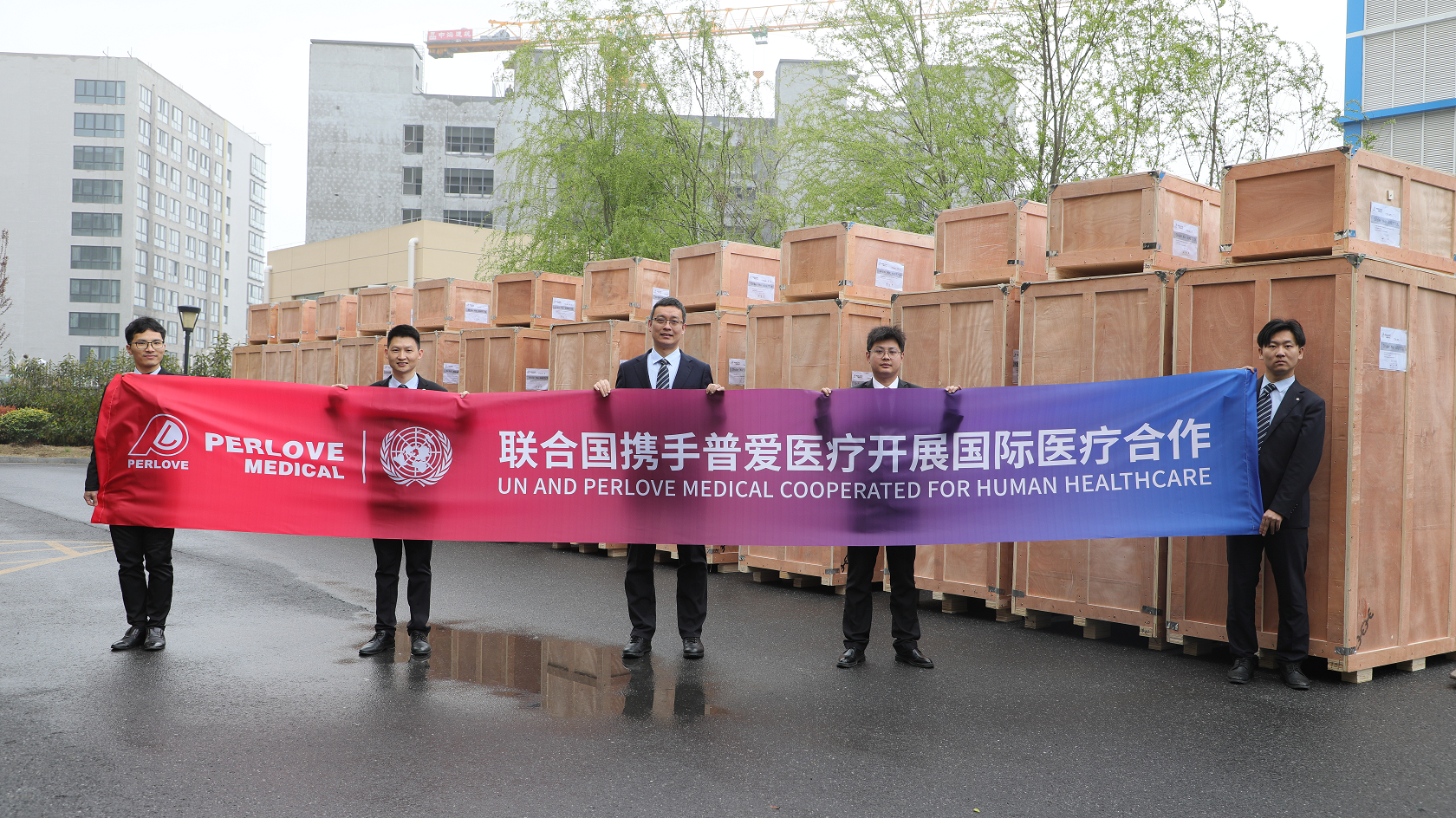
Read More »UN and Perlove Medical Cooperated for Human Healthcare
Read More »Perlove Medical Dynamic Flat DR injects new vitality into plateau medical services
Read More »China first!Perlove Medical's PLD9600 Series makes history as the first domestic DRF to obtain EU CE Certification
Read More »Perlove Medical's dynamic flat panel DRF successfully installed in Pakistan, Product quality proves reliability.
Read More »