The 2D C-arm has fluoroscopy and radiography, and can provide doctors with intraoperative image guidance, most widely used in orthopedics. While in recent years, the 3D C-arm is more and more popular. What is its unique advantage compared with 2D C-arm?
The 3D C-arm, also known as “intraoperative CT”, has all the functions of a 2D C-arm.In addition, it also has a 3D imaging function. The 3D C-arm rotates to collect 2D projection data from multiple angles, and then carries out 3D reconstruction through the computer software, which is capable of generating cross-sectional, sagittal, and coronal tomographic images as well as 3D stereoscopic images, providing more comprehensive 3D anatomical information.

2D AP/Lateral Views

3D "CT-like" Imaging
In clinical practice, the 3D C-arm reveals details that 2D imaging cannot capture:
Case 1: Osteopathy of the right distal femoral epiphysis (13 years old)

The lesion is not visible on 2D imaging

The 3D sectional images clearly reveal the lesion.
Case 2: Ankle Fracture Reduction (31 years old)
In ankle fracture reduction surgery, the reduction of the fracture site can be better visualized with 3D C-arm.

Good reduction of the tibiofibular syndesmosis on the AP and lateral views

3D imaging shows suboptimal reduction with inadequate congruency.
Case 3: Endobronchial Ultrasound-guided Transbronchial Lung Biopsy (80 years old)
The 3D C-arm can also be used innovatively in the field of respiratory intervention. For example, in scenarios such as percutaneous puncture or transbronchoscopic lung nodule biopsy, localization, and ablation, it is used to confirm that the tool reaches the lesion and that the ablation area covers the lesion completely.

2D imaging cannot accurately determine whether the instrument has reached the target lesion.

3D imaging confirms instrument placement within the target lesion.
There are inherent limitations in the images provided by 2D C-arms. Its projection-based principle captures only a single plane, resulting in the loss of depth information and frequent obscuration by overlapping bones or soft tissues. This makes certain lesions difficult to visualize or even completely hidden. In contrast, 3D C-arm can obtain CT-like tomography images through 3D reconstruction technology, thus solving the overlapping problem of 2D images.
Perlove Medical
With 3D images, doctors can perform more intuitive and in-depth analysis of pathologies, enabling comprehensive evaluation—such as precise assessment of fracture reduction, clear visualization of implant positioning, and spatial relationships with surrounding tissues. Particularly in complex clinical scenarios (e.g., complex fractures, spinal surgeries, and respiratory interventions), the detailed 3D data provided by 3D C-arms is critical for surgical navigation and outcome assessment.
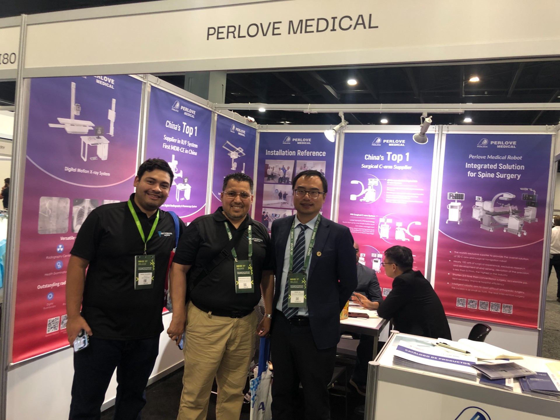
🌍 WHX Miami 2026 – Exhibition Wrap-Up
Read More »Perlove Medical to Exhibit at WHX Miami — See You in Miami!
Read More »A Decade of Dedication, Echoing in the Market Again | Perlove Medical’s C-arms Deployed in Three Major Regional Hospitals in Bangladesh
Read More »2026 Innovate Together, Grow Further丨Perlove Medical International Distributor Promotion Conference
Read More »PERLOVE MEDICAL at ECR 2026 | Advancing Radiology Innovation in Vienna
Read More »Perlove Medical at ECR 2026: Showcasing Cutting-Edge Imaging Solutions in Vienna
Read More »