PLX C2119
WIDER FIELD OF VIEW |  |
Simplicity revealed in every details |
Upgraded FPD |
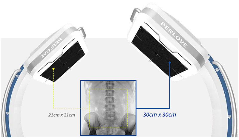
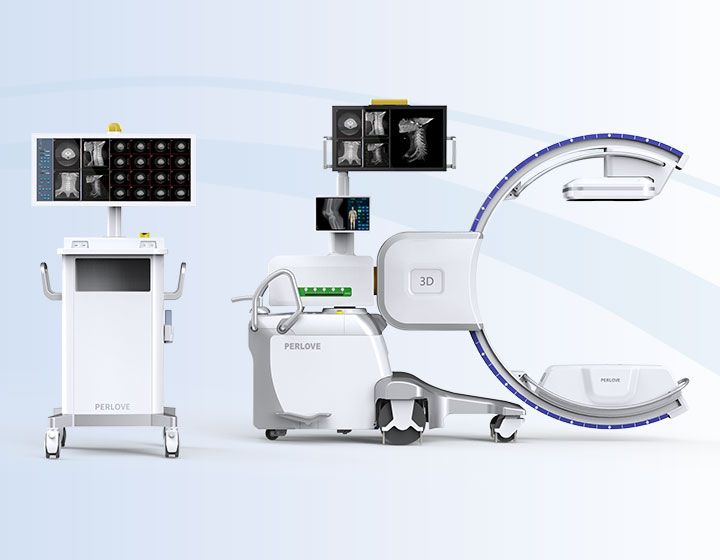
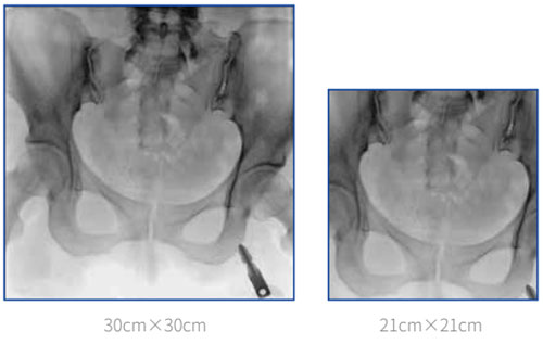
Larger field of view Equipped with a 30cm*30cm large-size dynamic FPD, imaging area has been increased by 100% compared with the conventional FPD.
Crystal-clear image with delicacy 4MP resolution image combined with low signal-noise ratio reveal the tiniest anatomical details. |
Maximize Effective AreaMinimize Exposure Time
5 lumbar vertebrae can be photographed within one image by a 30cm x 30cm FPD, largely decreasing exposure times caused by insufficient image FOV, making it easier for surgeons to locate lesions and to make surgical plans in the procedure like pedicle screw fixation or procedure with strict imaging demand. |
lmage taken by 30cm x 30cm FPD can basicly cover the entire pelvic floor. In the surgery of bilateral-pevic fractures or pelvic ring internal fixation, all the fracture sites can be revealed by single exposure, which significantly improves overall efficiency. |
Clinical image
Clinical application
Joint surgery lmage with high resolution can clearly reveal articular structure, helping surgeons to better locate the target. |
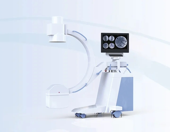
Trauma surgery Flexible mechanical movement realize easy positioning for different type of trauma surgery, providing comfortable handling experience.
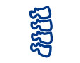
Spine surgery upgraded FPD present more details of vertebrae, helping surgeons to better making surgical plans.
Pain management Multiple radiation protection decrease overall exposure times during minimally invasive procedure.
|
Low dose management |
High frequency pulsed fluoroscopy X-ray generated in high frequency pulse wave can decrease 50% of radiation dose compared with a continous fluoroscopy. This technology has a positive effect on weaking the motion artifact.
DAP monitoring Real-time DAP displayed on the screen.
Intelligent parameter adjustment Optimized and adjustable exposure parameter created by intelligent hardware and algorithm provides a friendly examination scenario.
Virtual collimator Allowing radiologists to preview and to adjust exposure area before exposure, reducing unnecessary radiation.
Aluminum beam filtration Decreasing radiation level by absorbing soft X-ray. Ensuring high quality image for precise diagnosis and treatment. |
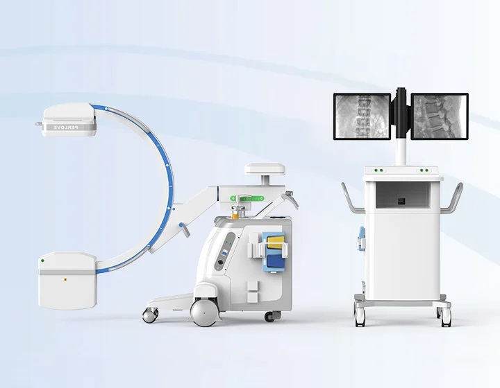
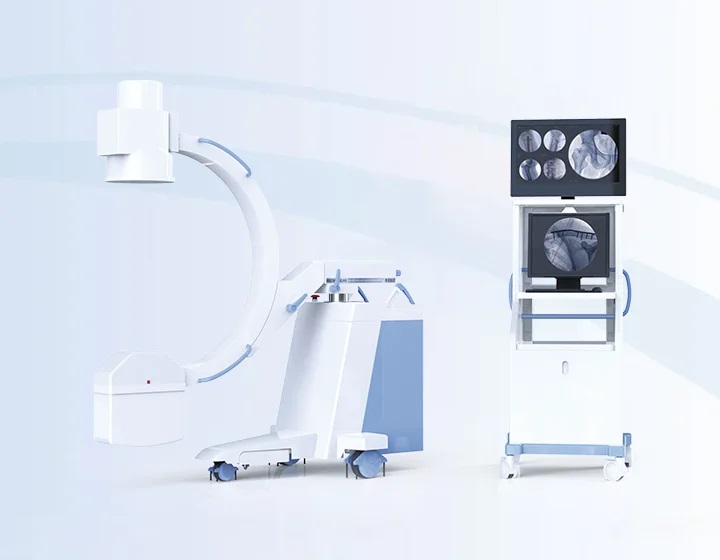
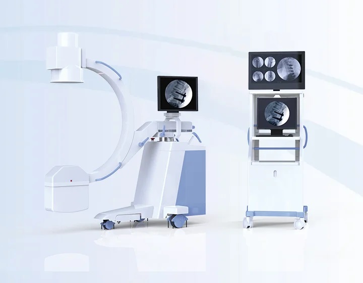
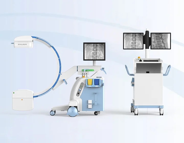
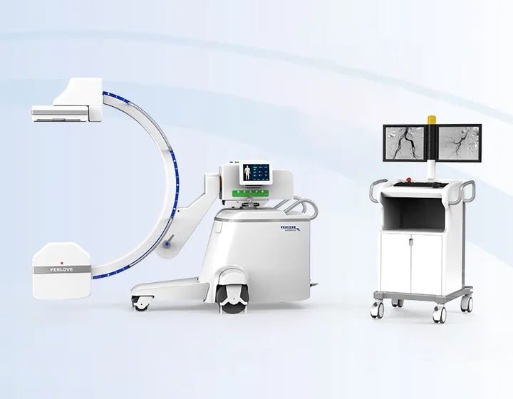
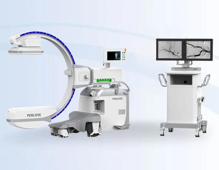
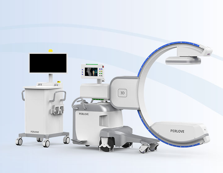
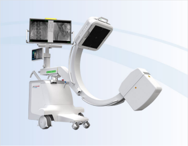
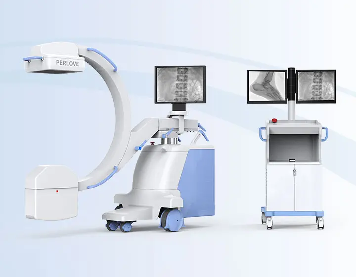
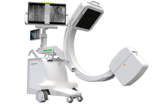
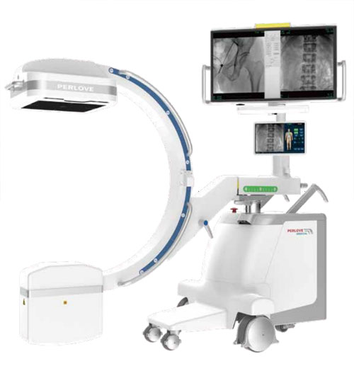
All-in-one Compact Design Flexible and EfficientCompact design + Standby power = A seamless connection among ORs. More convenient operation · The integrated workstation integrates the image acquisition system and the X-ray generator control system, which is convenient and quick to operate. Easy and flexible movement · The allin-one compact design of C-arm eliminates the monitor cart and wires, which makes it move flexibly. The machine can be easily carried out by operators with one hand. Instant surgery share · The machine is equipped with 10-minute standby power. When the power is cut off, the machine is able to remain activated and transfered to another OR without power connection. As soon as it is reconnected to the AC, the machine will immediately restore normal functions. | User-friendly Touch Interface
Easy exposure control Equipped with the high-quality and large-size touchscreen, high sensitivity, flexible rotation, more convenient for clinicians to perform touch operation. Large-size monitor Dual screen interface is convenient for surgeons to obtain more comprehensive clinical information Clear clinical images The sharp clinical images can be guaranteed. It can realize clear display of tiny anatomical structures, with no loss of image information, bringing more comfortable reading for surgeons. |


Larger field of view
Equipped with a 30cm*30cm large-size dynamic FPD, imaging area has been increased by 100% compared with the conventional FPD.

Crystal-clear image with delicacy
4MP resolution image combined with low signal-noise ratio reveal the tiniest anatomical details.

Full lumbar vertebrae imaging
5 lumbar vertebrae can be photographed within one image by a 30cm x 30cm FPD, largely decreasing exposure times caused by insufficient image FOV, making it easier for surgeons to locate lesions and to make surgical plans in the procedure like pedicle screw fixation or procedure with strict imaging demand.
Entire pelvic imaging
lmage taken by 30cm x 30cm FPD can basicly cover the entire pelvic floor. In the surgery of bilateral-pevic fractures or pelvic ring internal fixation, all the fracture sites can be revealed by single exposure, which significantly improves overall efficiency.


Joint surgery
lmage with high resolution can clearly reveal articular structure, helping surgeons to better locate the target.

Trauma surgery
Flexible mechanical movement realize easy positioning for different type of trauma surgery, providing comfortable handling experience.

Spine surgery
upgraded FPD present more details of vertebrae, helping surgeons to better making surgical plans.

Pain management
Multiple radiation protection decrease overall exposure times during minimally invasive procedure.


Lower leakage radiation in the loading state
Leakage radiation in the loading state of X-ray tube is far more lower than CFDA and FDA standard, providing better protection from the X-ray source.


High frequency pulsed fluoroscopy
X-ray generated in high frequency pulse wave can decrease 50% of radiation dose compared with a continous fluoroscopy. This technology has a positive effect on weaking the motion artifact.

DAP monitoring
Real-time DAP displayed on the screen.
Easy for monitoring radiation level.

Intelligent parameter adjustment
Optimized and adjustable exposure parameter created by intelligent hardware and algorithm provides a friendly examination scenario.

Virtual collimator
Allowing radiologists to preview and to adjust exposure area before exposure, reducing unnecessary radiation.

Aluminum beam filtration
Decreasing radiation level by absorbing soft X-ray. Ensuring high quality image for precise diagnosis and treatment.
Compact design + Standby power = A seamless connection among ORs.
More convenient operation
· The integrated workstation integrates the image acquisition system and the X-ray generator control system, which is convenient and quick to operate.
· When surgeons perform complex surgeries,it is easy for the surgeon to observe the surgical results and monitor the surgical process in time.
Easy and flexible movement
· The allin-one compact design of C-arm eliminates the monitor cart and wires, which makes it move flexibly. The machine can be easily carried out by operators with one hand.
Instant surgery share
· The machine is equipped with 10-minute standby power. When the power is cut off, the machine is able to remain activated and transfered to another OR without power connection. As soon as it is reconnected to the AC, the machine will immediately restore normal functions.

Easy exposure control
Equipped with the high-quality and large-size touchscreen, high sensitivity, flexible rotation, more convenient for clinicians to perform touch operation.
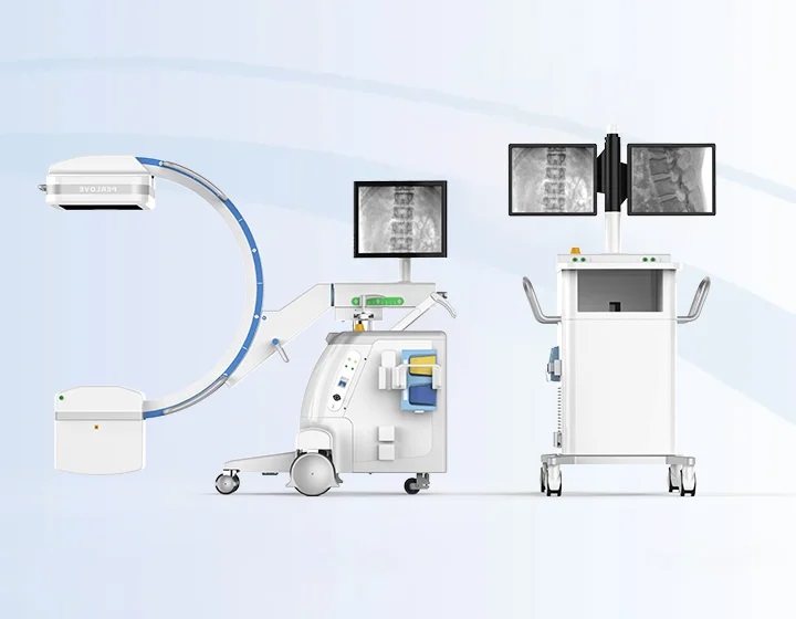
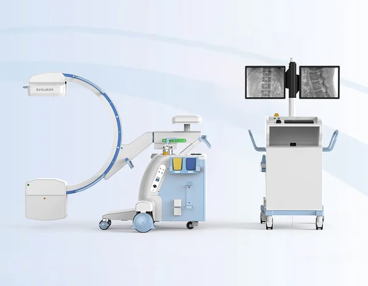
Large-size monitor
Dual screen interface is convenient for surgeons to obtain more comprehensive clinical information
Clear clinical images
The sharp clinical images can be guaranteed. It can realize clear display of tiny anatomical structures, with no loss of image information, bringing more comfortable reading for surgeons.